|
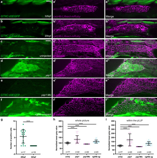
Yap1 transcriptional activity is detected in the pLLP prior to the onset of migration. a–b” Single-plane confocal images of the pLLP in control embryos at 32 hpf (a) and at 20 hpf (b) showing GTIIC:d2EGFP expression. cxcr4b:Lifeact-mRuby is used to visualize the pLLP (a’, b’). Merged images of the two channels are shown in (a”, b”). Single-plane confocal images of the pLLP in 20 hpf embryos (c), injected with tBFP-yap1 mRNA (d), yap1 Mo (e), or vgll4b sg1+2 (f), showing GTIIC:d2EGFP (c–f), β-catenin (c’–f’), and merged channels (c”-f”). β-catenin is used to visualize the pLLP. g Quantification of GTIIC:d2EGFP positive cells in the pLLP of 20 hpf and 32 hpf control embryos (N = 1 independent replicate). Mean gray value measured either on the whole image (h) or within the pLLP on a single plane (i) in the GFP channel in the same embryos for each condition (N ≥ 1 independent replicates). Data are presented as mean ± SD. Unpaired t-tests (Mann–Whitney) were conducted. Scale bar: 20 μm (a–f”).
|