|
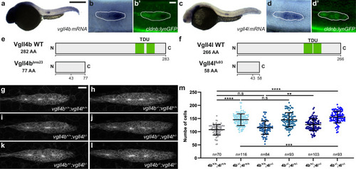
Vgll4b and Vgll4l are required to limit the number of cells in the pLLP. Overview (a, c) and higher magnification images (b, b’, d, d’) taken on a brightfield microscope of 32 hpf cldnb:lynGFP embryos stained by in situ hybridization (ISH) for vgll4b (a, b) and vgll4l (c, d) and counterstained with an anti-GFP antibody (b’, d’). Schematic representation of WT Vgll4b and the mutant form Vgll4bbns23 (e), and WT Vgll4l and the mutant form Vgll4lfu93 (f). MIP of confocal Z-stacks showing the pLLP in vgll4b+/+;vgll4l+/+ (g), vgll4b−/−;vgll4l+/+ (h), vgll4b+/+;vgll4l−/− (i), vgll4b−/−;vgll4l+/− (j), vgll4b+/−;vgll4l−/− (k), vgll4b−/−;vgll4l−/− (l) embryos. m Quantifications of pLLP cell number for each indicated group (N ≥ 4 independent replicates). Data are presented as mean ± SD. Unpaired t-tests (Mann–Whitney) were conducted. Scale bar: 400 μm (a, c), 20 μm (b–d’, g–l).
|