FIGURE SUMMARY
Title

Ddx3xa mutations drive cardiac defects in a zebrafish model via dysregulation of wnt/β-catenin signaling

Authors
Chen, Y., Lin, M., Zhu, P., Wang, H., Jiao, Z., Yi, K., Yang, X., Zhang, Y., Cai, X., Yuan, W., Li, Y., Jiang, Z., Wang, Y., Li, F., Wu, X., Fan, X.
Source
Full text @ Front Mol Biosci

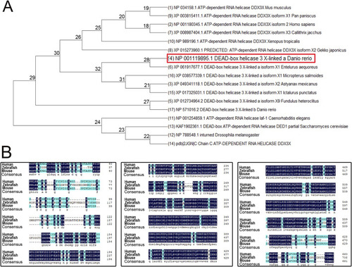
Phylogenetic tree and amino acid sequence analysis of DDX3X across species. (A) Phylogenetic tree of DDX3X homologs from diverse species. (B) Amino acid sequence alignment of Homo sapiens DDX3X, Mus musculus DDX3X, and Danio rerio Ddx3xa.

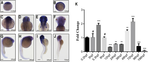
Spatiotemporal expression pattern of ddx3xa by whole-mount in situ hybridization and qRT-PCR analysis. (A–J) Spatiotemporal expression of ddx3xa at various developmental stages visualized by whole-mount in situ hybridization. Scale bar: 250 μm (applies to A–J). (A) 0.2 hpf; (B) 12 hpf; (C) 24 hpf; (D) 36 hpf; (E) 48 hpf; (F) 72 hpf; (G) Dorsal view, 24 hpf; (H) Dorsal view, 36 hpf; (I) Dorsal view, 48 hpf; (J) Dorsal view, 72 hpf. (K) Quantitative real-time PCR (qRT-PCR) analysis of relative ddx3xa transcript levels at various developmental time points (0.2, 3, 5.3, 8, 12, 24, 36, 48, 72, 96, 196 hpf). Expression levels were normalized to the 0.2 hpf time point. (n = 3; **P < 0.01, ***P < 0.001).

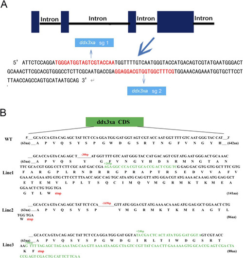
Schematic representation of ddx3xa knockout in zebrafish. (A) Design of sgRNAs targeting ddx3xa. Horizontal lines represent ddx3xa genomic DNA, blue rectangles denote exons, and red text indicates target site sequences. (B) Schematic of three heritable ddx3xa mutant alleles generated by CRISPR/Cas9 and their resultant protein sequences.

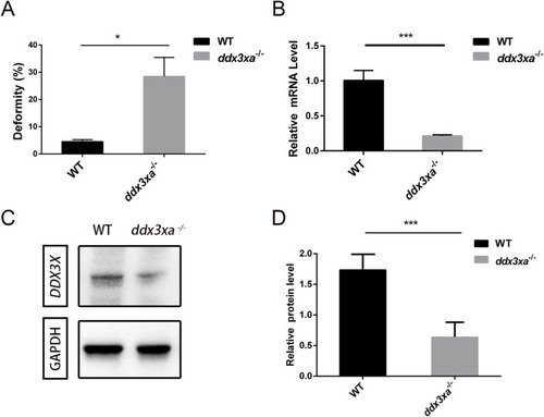
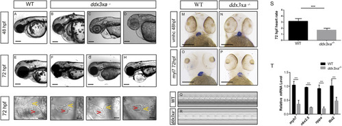
Malformation rate quantification and validation of ddx3xa knockout efficiency in zebrafish. (A) Malformation rate in ddx3xa−/− zebrafish at 48 hpf (**P < 0.01 vs. wild-type). (B) qRT-PCR analysis of ddx3xa mRNA levels in 72-hpf embryos showing significant downregulation in mutants (n = 3; *P < 0.05, ***P < 0.001). (C) Representative Western blot of Ddx3xa protein expression in 72-hpf larvae. Gapdh served as a loading control. (D) Quantitative densitometric analysis of Ddx3xa protein levels in wild-type and ddx3xa−/− larvae at 72 hpf.

EXPRESSION / LABELING:
Gene:
Fish:
Anatomical Term:
Stage: Protruding-mouth
PHENOTYPE:
Fish:
Observed In:
Stage Range: Long-pec to Protruding-mouth

ddx3xa knockout induces early cardiac malformation and dysfunction in zebrafish. (A–H) Phenotypic analysis of zebrafish hearts. (A) Normal cardiac phenotype in wild-type (WT) zebrafish at 48 hpf. (B–D) Cardiac phenotypes in ddx3xa−/− zebrafish at 48 hpf. (E) Normal cardiac phenotype in WT zebrafish at 72 hpf. (F–H) Cardiac phenotypes in ddx3xa−/− zebrafish at 72 hpf. Scale bar: 500 µm (applies to A–H). (I) Dorsal view of the heart in WT zebrafish at 72 hpf. (J–L) Dorsal views of hearts in ddx3xa−/− zebrafish at 72 hpf. Scale bar: 25 µm (applies to I–L). (M–N) Whole-mount in situ hybridization (WISH) with vmhc probe at 48 hpf. ddx3xa−/− larvae display irregular ventricular contours compared to WT (scale bar: 250 μm). (M)vmhc WISH in WT zebrafish at 48 hpf. (N) vmhc WISH in ddx3xa−/− zebrafish at 48 hpf. (O–P)myl7 probe WISH at 72 hpf. ddx3xa−/− larvae exhibit defective atrioventricular looping and atrial hypoplasia compared to WT (scale bar: 250 μm). (O)myl7 WISH in WT zebrafish at 72 hpf. (P)myl7 WISH in ddx3xa−/− zebrafish at 72 hpf. (Q–T) Heartbeat analysis in WT and ddx3xa−/− zebrafish at 72 hpf. (Q) Representative heartbeat trace of WT zebrafish at 72 hpf. (R) Representative heartbeat trace of ddx3xa−/− zebrafish at 72 hpf. (S) Statistical analysis of heart rate (n = 3; ***P < 0.001). (T) qRT-PCR analysis of myh7, nkx2.5, nppa, and tbx2 transcript levels in WT and ddx3xa−/− larvae at 72 hpf (n = 3; **P < 0.01, ***P < 0.001).

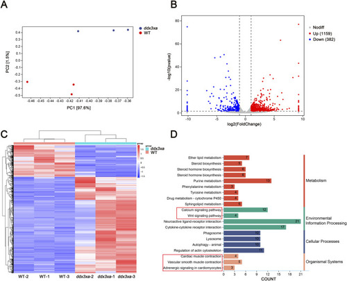
Transcriptomic alterations in ddx3xa−/− zebrafish at 72 hpf. (A) Principal component analysis (PCA) of the transcriptome revealed clear separation between WT and ddx3xa−/− groups along PC1, which accounted for 97.6% of the total variance (PC2: 1.5%). Sample distribution: wild-type (WT) controls (red) versus ddx3xa−/− mutants (blue). (B) Volcano plot of differentially expressed genes (DEGs): downregulated (blue), nonsignificant (gray), upregulated (red). (C) Hierarchical clustering heatmap: rows represent genes, columns represent samples. Expression levels scaled from high (red) to low (blue). (D) KEGG pathway enrichment analysis.

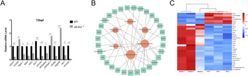
Validation and pathway analysis of cardiac development genes in ddx3xa−/− zebrafish. (A) qRT-PCR validation of genes related to cardiac contraction, vascular smooth muscle contraction, calcium signaling, and actin cytoskeleton regulation in ddx3xa−/− mutants versus wild-type (WT) at 72 hpf (n = 3; *P < 0.05, **P < 0.01, ***P < 0.001). (B) Pathway-gene interaction network for enriched cardiac development pathways generated using Cytoscape. (C) Heatmap of RNA-Seq expression profiles for enriched genes across key cardiac development pathways.

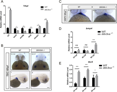
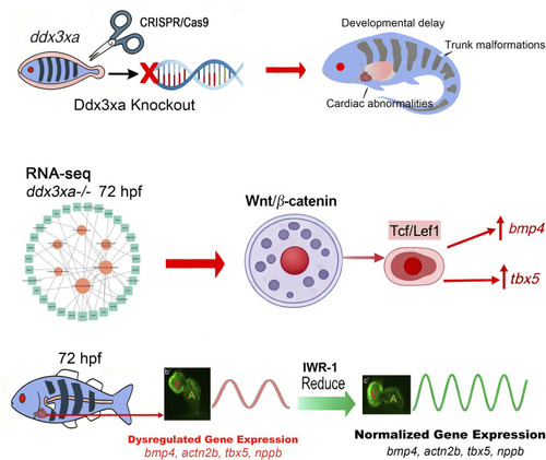
Wnt signaling disruption in ddx3xa−/− zebrafish. (A) Expression of key Wnt signaling components in ddx3xa−/− larvae at 72 hpf by qRT-PCR (n = 3; *p < 0.05, **p < 0.01, ***p < 0.001 vs. WT). (B) Whole-mount in situ hybridization (WISH) with tbx5 probe at 48 hpf. Numbers indicate identically stained embryos/total processed. Scale bar: 250 μm (applies to B–C). (a) WT dorsal view; (b) ddx3xa−/− dorsal view; (C) WT lateral view; (D)ddx3xa−/− lateral view. (C) WISH with bmp4 probe at 48 hpf. (a) WT ventral view; (b) ddx3xa−/− ventral view. (D)bmp4 expression levels in ddx3xa−/− embryos at 24, 48, and 96 hpf by qRT-PCR (n = 3; ***p < 0.001 vs. WT; ns: nonsignificant). (E)tbx5 expression levels in ddx3xa−/− embryos at 24, 48, and 96 hpf by qRT-PCR (n = 3; *p < 0.05, ***p < 0.001 vs. WT).

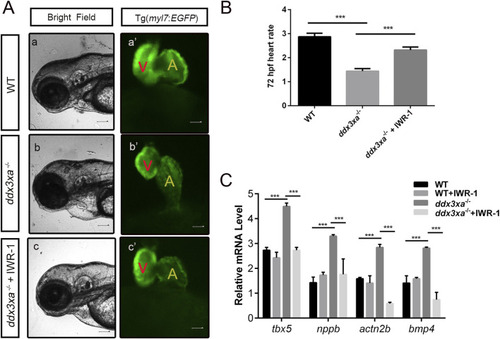
Pharmacological rescue of cardiac defects by Wnt inhibition. (A) Cardiac morphology at 72 hpf. Brightfield (A–C) and corresponding myl7:EGFP fluorescence (a'-c') views. (a,a') Wild-type (WT); (b,b') ddx3xa−/−; myl7:EGFP homozygous mutant; (c,c') ddx3xa−/−; myl7:EGFP mutant treated with IWR-1. Scale bar: 250 μm. (B) Heart rate quantification: ddx3xa−/− versus IWR-1-treated ddx3xa−/− mutants (n = 3; ***P < 0.001). (C) qRT-PCR analysis of Wnt-dependent cardiac genes (tbx5, nppb, actn2b, bmp4) in WT, WT + IWR-1, ddx3xa−/−, and ddx3xa−/− +IWR-1 embryos at 72 hpf (n = 3; ***P < 0.001).

ddx3xa Governs Cardiac Morphogenesis Through Wnt/β-Catenin Signaling. Schematic diagram illustrating the mechanistic role of ddx3xa in zebrafish cardiac development. CRISPR/Cas9-mediated ddx3xa knockout results in developmental defects including developmental delay, trunk malformations, and cardiac abnormalities characterized by pericardial edema and impaired looping. Transcriptomic profiling of ddx3xa−/− mutants at 72 hpf identifies dysregulation of key cardiac developmental genes (bmp4, actn2b, tbx5, nppb) and reveals hyperactivation of the Wnt/β-catenin pathway. Mechanistically, ddx3xa deficiency upregulates the transcription factor Tcf/Lef1, leading to aberrant expression of its downstream targets bmp4 and tbx5. Pharmacological inhibition of Wnt signaling with IWR-1 normalizes the expression of these cardiac genes and partially rescues the morphological defects. This work establishes ddx3xa as a critical regulator of cardiac development through modulation of the Wnt/β-catenin pathway, providing novel mechanistic insights into the cardiac comorbidities of DDX3X syndrome and highlighting the therapeutic potential of Wnt pathway modulation.

Acknowledgments
This image is the copyrighted work of the attributed author or publisher, and ZFIN has permission only to display this image to its users. Additional permissions should be obtained from the applicable author or publisher of the image. Full text @ Front Mol Biosci