FIGURE

Fig. 1

ID
ZDB-FIG-260805-21
Publication
Tian et al., 2026 - Regulation of the PKD2 channel function and associated disease phenotypes by RASSF4
Other Figures
All Figure Page
Back to All Figure Page
Fig. 1

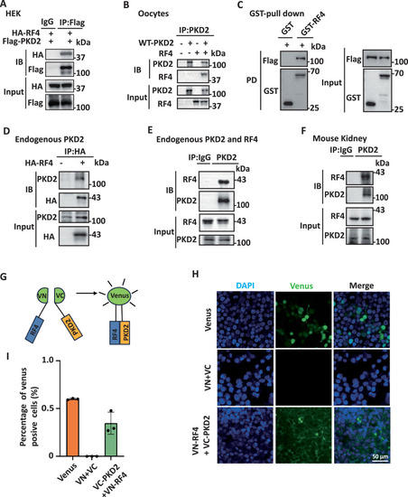
Physical interaction between PKD2 and RASSF4.
Representative data showing interaction between exogenous RASSF4 (RF4) and PKD2 in HEK cells (A) and Xenopus oocytes (B) detected using co-IP assays and WB. C Representative data showing interaction between purified GST-tagged RASSF4, expressed in E. coli BL21, and Flag-tagged PKD2, expressed in HEK293T cells, assessed by GST pull-down assays. Representative data showing interaction of over-expressed (D) and endogenous (E) RASSF4 with endogenous PKD2 in HEK cells, examined using co-IP. F Representative data showing interaction between endogenous RASSF4 and PKD2 assessed by co-IP in a kidney of an 8-week-old C57BL/6 mouse. G–I The RASSF4/PKD2 interaction was analyzed using BiFC assays. G Schematic presentation of a BiFC assay. The Venus N-terminal half (VN) and C-terminal half (VC) were tagged to the N-terminal end of RASSF4 and PKD2, respectively, for transfection in HEK cells. The green fluorescent signal indicates proximity between VN and VC, which indicates the presence of the RASSF4/PKD2 association. H Representative data showing fluorescence in HEK cells transfected with Venus, VN + VC or VN-RF4 + VC-PKD2. Nuclei were stained with DAPI. I Statistical data showing quantified fluorescence intensities obtained under the same conditions as in (H).

Expression Data

Expression Detail
Antibody Labeling
Phenotype Data

Phenotype Detail
Acknowledgments
This image is the copyrighted work of the attributed author or publisher, and ZFIN has permission only to display this image to its users. Additional permissions should be obtained from the applicable author or publisher of the image. Full text @ Commun Biol