FIGURE

Fig. 4

ID
ZDB-FIG-260805-24
Publication
Tian et al., 2026 - Regulation of the PKD2 channel function and associated disease phenotypes by RASSF4
Other Figures
All Figure Page
Back to All Figure Page
Fig. 4

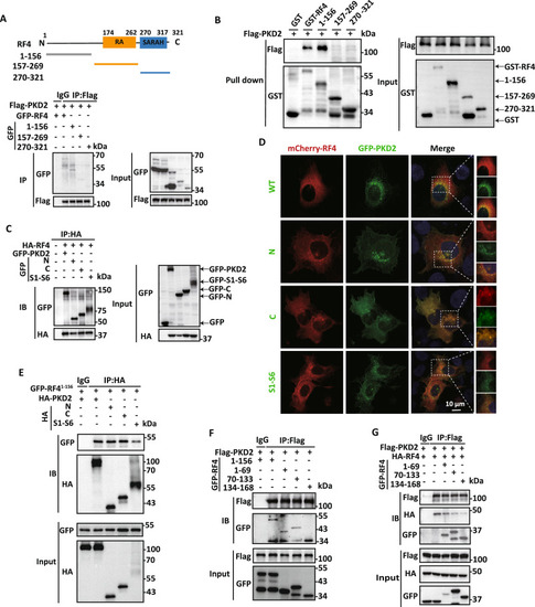
Domains mediating the RASSF4/PKD2 interaction.
A Upper panel, schematic architecture of human RASSF4 protein, with key functional domains indicated. Lower panels, representative co-IP data from HEK cells, showing interaction of Flag-PKD2 with GFP-RASSF4 and three GFP-tagged RASSF4 fragments. B Representative data obtained from GST pull-down assays showing the RASSF4/PKD2 interaction. HEK cells expressing Flag-PKD2 were incubated with purified GST-RASSF4 truncation proteins. Complexes were pulled down with glutathione beads and analyzed by WB. C Representative data from co-IP assays in HEK cells co-expressing HA-RASSF4 with different GFP-tagged PKD2 fragments (left panels) or co-expressing GFP-RASSF4 with different HA-tagged PKD2 fragments (right panels). “N”, PKD2 N-terminus (M1-S219), “C”, C-terminus (I671-V968), and “S1–S6”, central domain (K215-M675). D Representative confocal microscopy images showing subcellular colocalization in U-2 OS cells of mCherry-RASSF4 with different GFP-PKD2 constructs, as indicated. Scale bar: 10 μm. E Representative co-IP data showing interaction of GFP-tagged RASSF4-M1-P156 with different HA-PKD2 fragments, as indicated, in HEK cells. F, G Representative co-IP data showing interaction of Flag-PKD2 with different GFP-tagged RASSF4 fragments, as indicated, in HEK cells.

Expression Data

Expression Detail
Antibody Labeling
Phenotype Data

Phenotype Detail
Acknowledgments
This image is the copyrighted work of the attributed author or publisher, and ZFIN has permission only to display this image to its users. Additional permissions should be obtained from the applicable author or publisher of the image. Full text @ Commun Biol