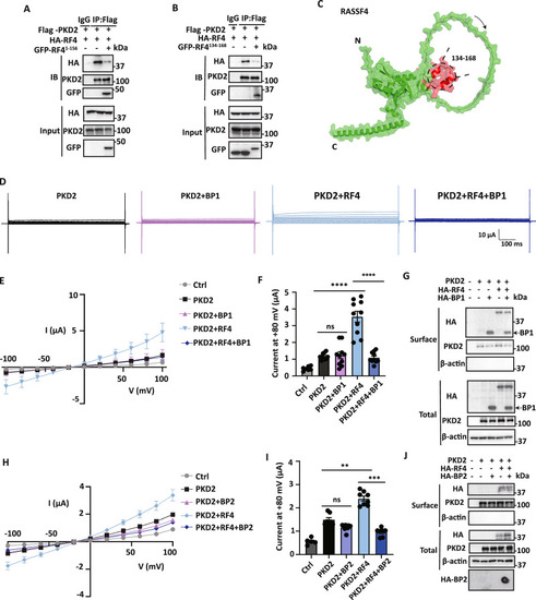
Effects of blocking peptides on the RASSF4/PKD2 binding and functional regulation of PKD2 by RASSF4.
The blocking effect of fragments M1-P156 (BP1) (A) and P134-S168 (BP2) (B) on the RASSF4/PKD2 interaction was shown by representative co-IP data obtained from HEK cells. C The Alphafold3 program predicted the presence of PKD2-binding, α-helical fragment in the RASSF4 N-terminus (P134-S168). D With similar electrophysiology conditions as in Fig. 1A, representative current traces obtained from oocytes expressing different constructs, as indicated, showed the blocking effect of BP1. E, F Averaged I–V curves and currents at +80 mV obtained under the same conditions as in (D), showing the blocking effect of BP1. Data are presented as the mean ± SD, with n = 7–10 oocytes for each condition. ****p < 0.0001, ns, not significant, by unpaired Student’s t test. G Representative biotinylation data showing the effect of HA-BP1 on the surface expression of PKD2 and RASSF4. H, I Averaged I–V curves and currents at + 80 mV obtained under similar conditions as in (D), showing the blocking effect of HA-BP2. n = 5–10 oocytes (H) and n = 5–10 oocytes (I) for each condition. Data are presented as mean ± SD. **p < 0.01, ***p < 0.001; ns, not significant, by unpaired Student’s t test. J Representative biotinylation data showing the effect of HA-BP2 on the surface expression of PKD2 and RASSF4. For dot blot analysis of HA-BP2, samples (2 µL) prepared by biotin labeling were spotted on a methanol-pretreated PVDF membrane, dried and UV-crosslinked. After blocking with 5% milk, BP2 was detected using an HA antibody. All subsequent steps were performed according to our WB protocol.
|
